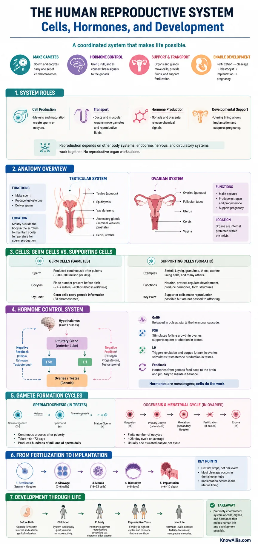
The human reproductive system is a coordinated set of organs, specialized cells, glands, ducts, and hormone signals that produces reproductive cells, supports fertilization, and makes human development possible. Its work depends on close communication between the brain, pituitary gland, ovaries or testes, reproductive tract, and developing tissues. The system is not active in exactly the same way throughout life; it forms before birth, remains relatively quiet through much of childhood, becomes active during puberty, and changes again with age.[a]
The System in Plain Terms
Human reproduction depends on three linked processes: making gametes, coordinating them with hormones, and supporting development after fertilization. Cells do the physical work, while hormones control timing, maturation, and feedback.
- Sperm and oocytes carry one set of 23 chromosomes each.
- GnRH, FSH, and LH connect brain signals to the gonads.
- Fertilization, cleavage, blastocyst formation, and implantation are separate steps rather than one event.
This article explains what the major reproductive organs do, how germ cells differ from supporting cells, how the hormone feedback system works, how gametes form, and how a fertilized cell develops through its earliest stages. It also separates several terms that are often treated as if they mean the same thing.
What the Reproductive System Does
The reproductive system has four broad biological jobs: it produces gametes, transports and supports those cells, produces reproductive hormones, and provides the conditions needed for fertilization and early development. The ovaries and testes are called gonads because they combine two roles that are often separated in other organ systems: they make cells and release hormones.[a]
- Cell production: meiosis and cell maturation create sperm or oocytes.
- Transport: ducts and muscular organs move gametes and reproductive fluids.
- Hormone production: gonadal and placental tissues release chemical signals.
- Developmental support: the uterine lining permits implantation and the uterus supports pregnancy.
Reproduction also depends on systems outside the reproductive organs. The endocrine system carries hormone messages through the blood, the nervous system helps control hormone release, and the circulatory system delivers oxygen, nutrients, and signaling molecules. No reproductive organ works alone.[l]
The Main Reproductive Organs
Human reproductive anatomy is commonly described as a testicular system and an ovarian system. Each has gonads, transport pathways, supporting glands or tissues, and external structures. Their parts differ, yet both systems solve the same biological problem: producing mature gametes and placing them where fertilization may occur.
| System | Structure | Primary Role |
|---|---|---|
| Testicular | Testes | Produce sperm within seminiferous tubules and release testosterone from Leydig cells. |
| Testicular | Epididymides | Support sperm maturation, concentration, storage, and transport. |
| Testicular | Vas deferens and ejaculatory ducts | Carry sperm toward the urethra. |
| Testicular | Seminal vesicles, prostate, and bulbourethral glands | Add fluids that form most of semen and support sperm transport. |
| Ovarian | Ovaries | Contain ovarian follicles, support oocyte maturation, and produce hormones including estradiol and progesterone. |
| Ovarian | Uterine tubes | Receive the ovulated oocyte and provide the usual site of fertilization. |
| Ovarian | Uterus | Builds a receptive lining for implantation and supports embryonic and fetal development. |
| Ovarian | Cervix and vagina | Connect the uterus with the exterior and form part of the pathway used by sperm, menstrual flow, and birth. |
| Ovarian | Vulva | Includes the external reproductive structures that protect openings and contain sensory tissues. |
The testes, ducts, accessory glands, and penis form the major parts of the testicular reproductive system.[b] The ovaries, uterine tubes, uterus, vagina, accessory glands, and external genital structures form the major parts of the ovarian reproductive system.[c]
The Cells That Make Reproduction Possible
The reproductive system contains two broad cell groups. Germ cells can enter the pathway that produces sperm or oocytes. Somatic supporting cells do not become gametes, but they shape the environment in which germ cells grow, divide, and respond to hormones.
Gametes: Sperm and Oocytes
Most nucleated somatic cells are diploid and contain 46 chromosomes. Mature gametes are haploid and contain 23 chromosomes. When a sperm nucleus and an oocyte nucleus combine at fertilization, the resulting zygote normally returns to the diploid number.[e]
Sperm Cell
- Head: contains the haploid nucleus.
- Acrosome: contains enzymes and proteins involved in interaction with the oocyte coverings.
- Midpiece: contains many mitochondria that supply energy.
- Flagellum: produces movement through fluid.
Oocyte
- Nucleus: carries the maternal chromosome set.
- Cytoplasm: contains organelles, proteins, and stored molecules used soon after fertilization.
- Zona pellucida: a specialized extracellular coat around the cell.
- Corona radiata: surrounding follicle cells that remain with the ovulated oocyte.
The oocyte is much larger than a sperm cell because it supplies most of the cytoplasm and organelles present in the newly formed zygote. Sperm are smaller and adapted for movement and delivery of a chromosome set. These are different cell designs for complementary tasks, not larger and smaller versions of the same cell.
Supporting Cells in the Gonads and Uterus
- Sertoli cells surround developing sperm cells, form a controlled local environment, respond to FSH, and release inhibin B.
- Leydig cells lie between seminiferous tubules and produce testosterone when stimulated by LH.
- Granulosa cells surround the developing oocyte, respond to FSH, and help produce estradiol.
- Theca cells respond mainly to LH and make androgen precursors used by granulosa cells.
- Luteal cells form after ovulation from the emptied follicle and produce progesterone along with estrogen.
- Endometrial cells build and remodel the uterine lining under changing estrogen and progesterone signals.
- Trophoblast cells form the outer cell population of the blastocyst and later contribute to the fetal portion of the placenta.
Supporting cells are not passive packaging. They regulate nutrients, local chemical signals, immune interactions, and the timing of cell maturation. In the testes, for example, sperm development depends on Sertoli cells as well as testosterone; in ovarian follicles, the oocyte and surrounding follicle cells exchange signals throughout maturation.
How Hormones Control the System
Reproductive hormone control is centered on the hypothalamic-pituitary-gonadal axis. The hypothalamus releases gonadotropin-releasing hormone, or GnRH, in pulses. GnRH acts on the anterior pituitary, which releases follicle-stimulating hormone and luteinizing hormone. FSH and LH then act on the ovaries or testes.[d]
The axis works less like a single on-off switch and more like a timed conversation. The brain sends a message, the pituitary relays it, the gonads respond, and hormones from the gonads report back. That feedback changes the next round of signals.
From Brain Signal to Reproductive Development
A linked sequence coordinates hormone release, gamete maturation, ovulation, sperm production, and preparation of reproductive tissues.
Hormone Signal Path
Releases GnRH in pulses rather than as a constant signal.
Responds by releasing FSH and LH into the bloodstream.
Produce gametes, steroid hormones, and inhibin in response to pituitary signals.
Estradiol, progesterone, testosterone, and inhibin alter further hormone release.
Cell-Level Results
Supports follicle growth and granulosa-cell activity.
Helps trigger ovulation and supports corpus luteum formation.
Acts through Sertoli cells to support sperm development.
Stimulates Leydig cells to produce testosterone.
Gamete Formation
Meiosis reduces chromosome number, while later cell changes produce a mature sperm or ovulated oocyte.
Cycle Timing
Hormone concentrations and tissue sensitivity change across the ovarian and uterine cycles.
Early Pregnancy
After implantation begins, trophoblast tissue produces hCG, which supports continued progesterone production early in pregnancy.
| Hormone | Main Source | Main Reproductive Actions |
|---|---|---|
| GnRH | Hypothalamus | Stimulates pituitary release of FSH and LH when delivered in pulses. |
| FSH | Anterior pituitary | Supports ovarian follicle growth and Sertoli-cell support of sperm development. |
| LH | Anterior pituitary | Triggers ovulatory events, supports corpus luteum formation, and stimulates testicular testosterone production. |
| Estradiol | Mainly ovarian follicles | Supports reproductive tissue growth, endometrial development, and feedback control of the brain and pituitary. |
| Progesterone | Corpus luteum and later placenta | Supports a secretory uterine lining and helps maintain conditions needed for pregnancy. |
| Testosterone | Mainly Leydig cells | Supports testicular development, sperm production, reproductive tissue function, and pubertal changes. |
| Inhibin | Sertoli or granulosa cells | Provides feedback that lowers FSH release. |
| AMH | Fetal Sertoli cells; granulosa cells later in life | Guides fetal reproductive-tract development in the testicular pathway; later reflects ovarian follicle activity. |
| hCG | Early trophoblast and placenta | Maintains corpus luteum hormone production during early pregnancy. |
| Prolactin | Anterior pituitary | Supports milk production after birth and interacts with reproductive hormone control. |
| Oxytocin | Made in the hypothalamus; released by the posterior pituitary | Supports uterine contractions and milk ejection. |
Hormones are often labeled “male” or “female,” but that wording is misleading. All people normally produce several of the same reproductive hormones. The differences lie mainly in concentration, timing, tissue source, receptor response, and life stage. Testosterone can be produced by ovaries and adrenal tissues, while estrogens are also formed in testicular and other tissues.
How Gametes Form and Mature
Spermatogenesis
Spermatogenesis takes place in the seminiferous tubules of the testes. Stem-like spermatogonia divide, selected cells enter meiosis, and the resulting haploid cells remodel into sperm. This remodeling includes nuclear condensation, formation of the acrosome, development of the flagellum, and removal of excess cytoplasm.
- Spermatogonia maintain the germ-cell population and produce cells that begin differentiation.
- Primary spermatocytes enter the first meiotic division.
- Secondary spermatocytes rapidly enter the second meiotic division.
- Spermatids are haploid cells that reshape through spermiogenesis.
- Spermatozoa are released from Sertoli-cell support and continue maturing in the epididymis.
FSH acts mainly through Sertoli cells, while LH stimulates testosterone production by Leydig cells. High local testosterone within the testes and Sertoli-cell support are both needed for normal sperm formation.
Oogenesis and Follicle Development
Oogenesis begins before birth. Developing germ cells become primary oocytes and pause in the first meiotic division. From puberty onward, groups of follicles may begin growing during each cycle, though usually only one becomes the dominant ovulatory follicle. Shortly before ovulation, the selected oocyte completes the first meiotic division and becomes a secondary oocyte. The second meiotic division is completed only if fertilization activates the cell.[f]
Follicle development is not simply an egg “waiting in storage.” A follicle is a living unit made of an oocyte and several layers of supporting cells. Those cells respond to hormones, produce local signals, and prepare the oocyte for ovulation.
The Ovarian and Uterine Cycles
The ovarian cycle and uterine cycle occur together, but they describe different tissues. The ovarian cycle follows follicle growth, ovulation, and corpus luteum activity. The uterine cycle follows changes in the endometrium.
| Phase | Ovary | Uterus | Dominant Hormone Pattern |
|---|---|---|---|
| Early Cycle | Several follicles begin growing under FSH influence. | Menstruation sheds part of the previous functional lining. | Estrogen and progesterone are relatively low at the start. |
| Follicular / Proliferative | A dominant follicle develops and produces more estradiol. | Estradiol supports rebuilding and thickening of the endometrium. | Estradiol rises; FSH is moderated by feedback. |
| Ovulation | An LH surge helps release the secondary oocyte. | The lining remains prepared rather than being shed. | LH rises sharply after sustained high estradiol feedback. |
| Luteal / Secretory | The emptied follicle becomes the corpus luteum. | Progesterone promotes a secretory, implantation-ready lining. | Progesterone is prominent, with estrogen also present. |
| Cycle Reset Without Pregnancy | The corpus luteum regresses. | Falling hormones lead to breakdown and shedding of the functional lining. | Progesterone and estrogen fall. |
A 28-day cycle is a useful teaching model, not a rule. Adult cycles commonly vary, and the day of ovulation is not fixed at day 14. Cycle length can differ between people and from one cycle to another.[g]
From Fertilization to Implantation
Fertilization usually occurs in the widened part of a uterine tube called the ampulla. Before a sperm can fuse with an oocyte, it undergoes capacitation, a set of changes that occurs within the reproductive tract. The sperm then interacts with the cells around the oocyte and the zona pellucida.
- Recognition and binding: sperm contacts the oocyte coverings through species-specific molecular interactions.
- Membrane fusion: one sperm fuses with the oocyte membrane and delivers its genetic material.
- Block to additional sperm entry: the oocyte changes its outer environment to reduce entry by other sperm.
- Completion of meiosis: the secondary oocyte completes meiosis II.
- Pronuclear union: the two haploid chromosome sets come together in the new zygote.
Fertilization creates a one-cell zygote, but implantation has not yet happened. The zygote divides by mitosis as it moves toward the uterus. These early divisions are called cleavage because cell number rises while the total conceptus remains within the zona pellucida.
Fertilization and pregnancy testing occur at different points. A pregnancy test detects hCG after trophoblast tissue begins producing enough hormone following early implantation. It does not detect the instant when sperm and oocyte fuse.[m]
| Stage | What Happens | Approximate Timing |
|---|---|---|
| Zygote | A single diploid cell forms after fertilization. | Day 0 |
| Cleavage | Mitotic divisions produce smaller cells called blastomeres. | Days 1–3 |
| Morula | A compact ball of cells forms as cell-to-cell contact changes. | About days 3–4 |
| Blastocyst | A fluid-filled structure forms with an inner cell mass and outer trophoblast. | About day 5 |
| Hatching | The blastocyst emerges from the zona pellucida. | Usually before implantation |
| Implantation | The blastocyst attaches to and begins entering the receptive endometrium. | Begins around the transition into the second week |
By the blastocyst stage, the inner cell mass will give rise to the embryo proper, while the trophoblast contributes to placental tissues. Implantation requires coordination between the developmental state of the blastocyst and the hormone-prepared endometrium.[i]
How the Reproductive System Develops
Reproductive development begins well before puberty. Early embryos form bipotential gonadal tissue, meaning the same early tissue can follow an ovarian or testicular developmental pathway. The internal ducts and external structures also begin from early tissues that later differentiate under genetic and hormone signals.[h]
Fetal Reproductive Development
In a typical testicular pathway, gene activity associated with testis development leads to formation of Sertoli and Leydig cells. Sertoli cells release anti-Müllerian hormone, while Leydig cells produce testosterone. These signals guide development of the internal reproductive tract and external structures. In a typical ovarian pathway, a different gene program supports ovarian development, and the absence of fetal testicular hormone signals permits Müllerian structures to develop into the uterine tubes, uterus, and upper portion of the vagina.[k]
Chromosomes, genes, hormone production, hormone conversion, receptors, and tissue timing all contribute to reproductive differentiation. A single label cannot describe every step. The process is a sequence of linked cellular decisions.
Puberty and Maturation
During much of childhood, the reproductive hormone axis is relatively quiet. Puberty begins when pulsatile GnRH activity rises and the pituitary increases LH and FSH release. The gonads become more active, gamete maturation advances, and reproductive tissues respond to higher steroid-hormone exposure.
- In ovarian development, early visible changes usually begin before the first menstrual period.
- In testicular development, enlargement of the testes is generally an early physical change.
- Growth, body composition, skin, hair, voice, bone, and reproductive organs can all change during puberty.
- The timing and pace vary. MedlinePlus describes common starting ranges of about ages 8–13 for girls and 9–14 for boys.[j]
Puberty is a process, not a single birthday or event. Menarche does not mean that every cycle will immediately include ovulation, and the first appearance of sperm production does not mean the entire system has reached its later adult pattern.
Changes Across Adult Life
Reproductive physiology continues to change after puberty. Ovarian follicle number declines with age, cycles may become less regular during the transition toward menopause, and menopause marks the permanent end of menstrual cycles after ovarian hormone patterns change. Testicular sperm production may continue into later life, although hormone output, semen characteristics, and fertility can change gradually rather than ending at one fixed point.[n]
Common Confusions About Reproductive Biology
Essential Terms
- Gamete
- A haploid reproductive cell: sperm or oocyte.
- Gonad
- An ovary or testis; an organ that produces gametes and reproductive hormones.
- Meiosis
- A specialized cell-division process that reduces chromosome number and creates genetic variation.
- Gametogenesis
- The formation and maturation of sperm or oocytes.
- Follicle
- An ovarian unit containing an oocyte and surrounding supporting cells.
- Ovulation
- Release of a secondary oocyte from a mature ovarian follicle.
- Corpus Luteum
- A temporary endocrine structure formed from the ovulated follicle.
- Endometrium
- The inner uterine lining that changes across the cycle and receives an implanting blastocyst.
- Fertilization
- Fusion-related events that combine sperm and oocyte genetic material to form a zygote.
- Cleavage
- Early mitotic divisions of the zygote that increase cell number.
- Blastocyst
- An early fluid-filled developmental stage with an inner cell mass and trophoblast.
- Implantation
- The process by which the blastocyst attaches to and enters the endometrium.
- HPG Axis
- The hormone communication pathway linking the hypothalamus, pituitary, and gonads.
- Feedback
- A control process in which hormone output changes earlier signals in the same system.
What Biology Textbooks Cannot Show Fully
Diagrams and phase tables simplify a system that changes from hour to hour and differs among individuals. Hormone concentrations are pulsatile or cyclic, tissue sensitivity can change, and one blood measurement may not describe an entire cycle. Exact timing of ovulation, fertilization, implantation, puberty, and reproductive aging cannot be predicted from a general diagram alone.
Several early events are also difficult to observe directly in an unassisted human pregnancy. Much of the detailed timing used in textbooks comes from clinical observation, laboratory research, imaging, hormone measurements, and assisted-reproduction settings. These sources provide useful models, but individual development does not follow a stopwatch.
Personal symptoms, cycle changes, puberty concerns, fertility questions, or pregnancy questions require individual medical assessment. General anatomy and physiology explain how the system usually works; they do not diagnose a specific person.
Frequently Asked Questions
What is the main function of the human reproductive system?
Its main biological functions are to produce gametes and reproductive hormones, transport and support gametes, allow fertilization, and support development after implantation.
Why do sperm and oocytes have only 23 chromosomes?
Meiosis reduces the chromosome number by half. This allows the chromosome sets from sperm and oocyte to combine at fertilization without doubling the usual human chromosome number in every generation.
What is the difference between an egg and an oocyte?
In everyday language, “egg” is often used for several stages. In precise biology, an oocyte is the developing female germ cell. The cell released at ovulation is usually a secondary oocyte, and it completes meiosis II only after fertilization begins.
Which hormones start the reproductive hormone pathway?
GnRH from the hypothalamus stimulates the anterior pituitary to release FSH and LH. These hormones act on the ovaries or testes, which then produce gametes, steroid hormones, and inhibin.
Does ovulation always happen on day 14?
No. Day 14 applies only as an approximate midpoint in a textbook 28-day cycle. Real cycle lengths vary, and ovulation timing varies with them.
Where does fertilization usually happen?
Fertilization usually occurs in the ampulla, the wider portion of a uterine tube. The resulting early conceptus then travels toward the uterus.
Are fertilization and implantation the same event?
No. Fertilization forms a zygote. Implantation occurs several days later, after cleavage and blastocyst formation, when the blastocyst begins attaching to the uterine lining.
What does hCG do in early pregnancy?
hCG is produced by early trophoblast tissue. It supports the corpus luteum so progesterone production can continue while early placental development proceeds.
Why are supporting cells important?
Germ cells cannot mature normally without them. Sertoli, Leydig, granulosa, theca, luteal, endometrial, and trophoblast cells provide hormones, nutrients, local signals, tissue structure, and controlled environments.
Do only males produce testosterone and only females produce estrogen?
No. All people normally produce and use androgens and estrogens. Typical differences involve the amount produced, the main tissue source, timing, and how target tissues respond.
Sources
- [a] ↩ National Cancer Institute SEER Training – Introduction to the Reproductive System — Primary system functions and the role of gonads.
- [b] ↩ National Cancer Institute SEER Training – Male Reproductive System — Testes, ducts, accessory glands, and external structures.
- [c] ↩ National Cancer Institute SEER Training – Female Reproductive System — Ovaries, uterine tubes, uterus, vagina, and external structures.
- [d] ↩ NCBI Bookshelf – The Hypothalamic-Pituitary-Gonadal Axis — GnRH, FSH, LH, gonadal function, and hormone feedback.
- [e] ↩ OpenStax Biology 2e – Human Reproductive Anatomy and Gametogenesis — Gametes, reproductive anatomy, Sertoli cells, Leydig cells, and sperm development.
- [f] ↩ NCBI Bookshelf – Oogenesis — Oocyte formation, meiotic arrest, and the cellular features of the oocyte.
- [g] ↩ NICHD – Menstruation Fact Sheet — Menstrual-cycle phases, cycle-length variation, and ovulation timing.
- [h] ↩ Endotext – Sexual Differentiation — Fetal gonadal, duct, and external reproductive development.
- [i] ↩ NCBI Bookshelf – Embryology, Week 1 — Fertilization, cleavage, morula, blastocyst formation, and implantation.
- [j] ↩ MedlinePlus – Puberty — Typical puberty timing ranges and common physical changes.
- [k] ↩ OpenStax Anatomy and Physiology 2e – Development of the Reproductive Systems — Bipotential tissues, fetal hormone signaling, and puberty-related maturation.
- [l] ↩ MedlinePlus – Hormones — Hormones as bloodstream-borne chemical messengers involved in growth, development, and reproduction.
- [m] ↩ Endotext – Endocrinology of Pregnancy — Implantation-related signaling, hCG production, and corpus luteum support.
- [n] ↩ NICHD – About Menopause — Hormone changes, cycle transition, and the definition of menopause.