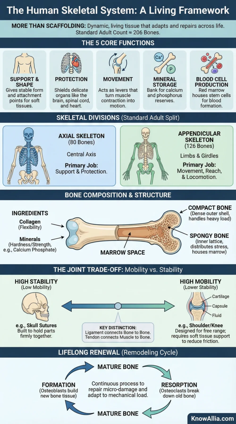
The human skeletal system is the body’s internal support system: a connected set of bones, joints, cartilage, and ligaments that gives shape, protects organs, stores minerals, and helps muscles turn effort into movement. In a typical adult, it is described as 206 named bones, but the bigger point is function: the skeleton is not dead support material. It is living tissue that changes, repairs, and adapts across life.[b][a][f]
What Matters Most
Bones, joints, and connective tissues work as one mechanical system. Bones provide shape and leverage, joints decide where motion can happen, and cartilage, ligaments, and marrow make that system usable, cushioned, and biologically active.[d][h]
- The adult skeleton is usually divided into 80 axial bones and 126 appendicular bones.[b]
- Bone tissue is made from collagen plus mineral, with dense outer layers and lighter inner trabecular regions.[a][g]
- Joints range from fixed skull sutures to freely movable synovial joints such as the knee and shoulder.[c]
This article follows the skeleton from its broad layout to bone tissue, joint design, renewal across life, and the places where simple textbook summaries can blur what is really happening inside the body.
What the Skeleton Actually Does
Most overviews stop at “support and movement,” but the skeleton does more than hold the body up. It protects delicate organs, acts as a mineral reserve, and houses marrow where blood cell formation begins. That is why the skeleton belongs in biology, not only in basic anatomy charts.[f][h]
- Support and Body Shape: bones give the body stable form and provide attachment points for soft tissues.[b]
- Protection: the skull surrounds the brain, the vertebral column protects the spinal cord, and the rib cage shields the heart and lungs.[b]
- Movement: muscles pull on bones, and bones act as levers that turn muscle force into visible motion.[b]
- Mineral Balance: bone stores calcium and phosphorus and can release them when the body needs them elsewhere.[f]
- Blood Cell Production: red marrow contains blood stem cells that can become red cells, white cells, and platelets.[h]
- Support
- Protection
- Movement
- Mineral Storage
- Blood Cell Formation
A useful mental model is this: the skeleton is not a rigid prop holding the body still. It is a load-bearing system that stays biologically active while you stand, walk, lift, heal, and grow.
How the Skeleton Is Arranged
The adult skeleton is usually divided into two large sections. The axial skeleton forms the central line of the body, while the appendicular skeleton includes the limbs and the girdles that attach them.[b]
Axial Skeleton
80 bones arranged around the head, neck, chest, and spine. This section is mostly about support and protection of the brain, spinal cord, and thoracic organs.[b]
- Skull
- Vertebral Column
- Ribs
- Sternum
Appendicular Skeleton
126 bones in the upper limbs, lower limbs, shoulder girdles, and pelvic girdle. This section is mostly about reach, grip, stance, and locomotion.[b]
- Shoulder Girdles
- Arms and Hands
- Pelvic Girdle
- Legs and Feet
| Division | Named Bones | Main Regions | Main Functional Emphasis |
|---|---|---|---|
| Axial | 80 | Skull, vertebral column, ribs, sternum | Support, organ protection, central body alignment |
| Appendicular | 126 | Shoulders, arms, hands, pelvis, legs, feet | Movement, reach, balance, transfer of body weight |
This split is easy to remember, but it also explains function. The axial section stabilizes the body’s middle line; the appendicular section gives the body range, stride, grip, and directional movement.
What Bone Tissue Is Made Of
Bone feels hard because it is mineralized, but it is not made of mineral alone. Bone contains collagen and minerals, especially calcium phosphate. The mineral contributes hardness and strength, while collagen adds enough flexibility to help bone resist breaking under force.[a]
Each bone also has an outer and inner design. The dense outer shell is compact or cortical bone. The inner lattice-like region is spongy, cancellous, or trabecular bone. These are not “better” and “worse” versions of bone; they serve different jobs inside the same organ.[g]
Spongy Bone
Lighter and built around a network of trabeculae. It helps distribute stress and leaves space for marrow within many bones.[g]
Bone Marrow
Soft tissue inside many bones. Red marrow contains stem cells that can become blood cells, while yellow marrow is richer in fat.[h]
In long bones, this design becomes especially clear: a strong shaft, widened ends, a marrow space, and articular surfaces where joints form. That arrangement gives the body a smart mix of strength, reduced weight, and biological activity.[k]
How Bones and Joints Keep the Body Working
The skeleton is useful because hard tissue, softer connective tissue, and controlled motion all work together at the same time.
Shape and Support
Bones define body form and give soft tissues stable attachment points. Without that support, posture and efficient movement would collapse.
Protection
Flat and curved bones around the skull and chest create protective spaces for the brain, heart, and lungs.
Controlled Motion
Joints decide where movement is allowed, how wide that movement can be, and how much stability must be preserved.
Mineral Reserve
Bone stores calcium and phosphorus, which helps the body balance mineral needs beyond the skeleton itself.
Compact Bone
Dense outer tissue that handles load and resists bending.
Trabecular Bone
Inner lattice that spreads stress and creates room for marrow.
Marrow
Soft tissue inside bone where blood-forming cells are found in red marrow.
The Main Bone Shapes
Bone shape is not random. A common basic classification groups bones into four main shape categories: long, short, flat, and irregular. Shape often hints at job, though real bones can do more than one thing at once.[j]
| Bone Type | Typical Form | What That Form Helps With | Examples |
|---|---|---|---|
| Long | Longer than wide, with a shaft and ends | Leverage and broad-range limb movement | Femur, humerus, radius, tibia |
| Short | Roughly cube-shaped | Stability with modest motion | Carpal and tarsal bones |
| Flat | Thin, flattened, often curved | Protection and wide muscle attachment surfaces | Many skull bones, sternum, ribs |
| Irregular | More complex shapes | Specialized support, passageways, and attachments | Vertebrae, some skull bones |
That is why a femur does not look like a vertebra and a vertebra does not look like a rib. Form follows job, and bone anatomy reflects the loads, attachments, and motions each region must handle.
How Joints Allow Motion Without Bone-on-Bone Contact
A joint, also called an articulation, is the place where two bones meet. Joints can be classified by what they are made of and by how much movement they allow. That is a better way to understand joints than treating them all as versions of the knee or elbow.[c]
In freely movable joints, the bone ends are covered with articular cartilage and separated by a joint space. The whole area is enclosed by a capsule, supported by ligaments, and lined by tissues that help maintain smooth motion. In other words, a healthy joint is designed to reduce direct friction, not to let bare bone grind on bare bone.[c][d][e]
A synovial joint works a bit like a well-fitted door hinge wrapped in a soft sleeve and supplied with a thin lubricant film: motion is guided, weight is transferred, and wear is kept under control. The analogy is not exact, but it helps explain why joint anatomy includes more than two touching bones.
| Joint Class | Main Tissue Feature | Usual Movement | Typical Example |
|---|---|---|---|
| Fibrous | Bones joined by fibrous connective tissue | Usually fixed or nearly fixed | Skull sutures |
| Cartilaginous | Bones linked by cartilage | Slight movement | Pubic symphysis, joints between vertebrae |
| Synovial | Joint cavity, cartilage-covered surfaces, capsule | Free movement | Knee, shoulder, hip, elbow |
Stability First
Some joints are built mainly to hold parts together. Skull sutures barely move, and cartilaginous joints such as those between vertebrae allow only limited motion because stability is part of the job.[c]
This stability-versus-mobility trade-off is one of the most useful ways to read joint anatomy. The body does not give every joint the same freedom because each region is solving a different mechanical problem.
How Bones Grow, Repair, and Renew Themselves
Bone is often described as if it were fixed once adulthood arrives. That is not how it works. Bone development begins early, continues through growth, and remains active later for repair and remodeling.[i]
Two words help here. Modeling changes the size and shape of bones during growth. Remodeling replaces older bone with newer bone at the same site across life. By the early adult years, remodeling becomes the dominant pattern, and it continues throughout life.[f]
- Formation: osteoblasts build bone tissue.[g]
- Maturity: osteocytes are mature bone cells embedded in bone matrix.[g]
- Resorption: osteoclasts break down and reabsorb bone as part of renewal.[g][i]
- Repair and Adaptation: the balance among these cells changes when bones heal after injury or respond to altered mechanical load.[f][i]
Development also follows two broad pathways. Intramembranous ossification forms certain flat skull bones and some irregular bones. Endochondral ossification forms most of the skeleton by replacing a cartilage model with bone tissue.[i]
One detail many basic articles miss is that bone remodeling is tied to whole-body physiology, not only to shape. Bone acts as a mineral reserve, and the body can draw on that reserve when calcium or phosphorus is needed elsewhere. That makes bone a structural organ and a metabolic organ at the same time.[f]
Age changes matter here too. Bone mass can decline over time, vertebrae can thin, and the discs between vertebrae can lose fluid and become thinner. Those changes help explain why posture, height, and joint stiffness may shift with age.[e]
Where People Often Get Mixed Up
Several common mix-ups make the skeletal system seem flatter and simpler than it really is. Clearing them up makes the whole topic easier to read.
Terms Worth Knowing
- Bone Marrow
- Soft tissue inside many bones. Red marrow contains blood-forming stem cells; yellow marrow contains more fat.[h]
- Cartilage
- Smooth supportive tissue found in several places, including the surfaces of movable joints where it cushions contact.[d]
- Ligament
- Fibrous connective tissue that connects bone to bone and helps hold structures together.[l]
- Tendon
- Fibrous connective tissue that connects muscle to bone and helps transmit muscle force.[l]
- Compact Bone
- The denser outer part of bone tissue, built for strength and load handling.[a][g]
- Spongy Bone
- The lighter inner trabecular region found in many bones, often around marrow spaces.[a][g]
- Ossification
- The process by which bone tissue forms during growth and development.[i]
- Remodeling
- The ongoing replacement of older bone with newer bone at the same site across life.[f]
- Synovial Joint
- A freely movable joint with a cavity, cartilage-covered surfaces, and a capsule.[c]
- Articular Cartilage
- Hyaline cartilage covering the ends of bones in freely movable joints.[c]
Where the Usual Numbers Need Context
The adult count of 206 bones is the standard named count used in anatomy. It is accurate as a teaching norm, but it is not perfectly fixed in every person. Bone number can vary because some bones fuse during development and because anatomical variation can change what gets counted in a given body.[k]
The same caution applies to simple charts of joints and bone types. They are useful maps, but real anatomy includes overlap, variation, and regional detail that short summaries often leave out.
FAQ
Questions Readers Often Ask
What Is the Main Job of the Skeletal System?
How Many Bones Are in the Adult Human Body?
The standard adult count is 206 named bones, divided into the axial and appendicular skeleton.[b]
Are Bones Living Tissue?
What Is the Difference Between a Bone and a Joint?
Why Can the Shoulder Move More Than the Skull?
Because different joints are built for different jobs. Skull sutures are built mainly for stability, while synovial joints such as the shoulder are designed for freer movement.[c]
What Is the Difference Between a Tendon and a Ligament?
A tendon connects muscle to bone. A ligament connects bone to bone and usually helps stabilize a joint.[l]
Sources
- [a] NIAMS – What Is Bone? — Bone composition, collagen, minerals, and the compact-versus-spongy tissue distinction.
- [b] NCI SEER Training – Divisions of the Skeleton — Adult bone count, axial skeleton, and appendicular skeleton.
- [c] NCBI Bookshelf – Anatomy, Joints — Joint definition, structural classes, and movement-based classes.
- [d] MedlinePlus – The Structure of a Joint — Joint components such as cartilage, ligaments, synovium, and load-bearing design.
- [e] MedlinePlus – Aging Changes in the Bones, Muscles, and Joints — Normal age-related changes in bone density, vertebrae, discs, and joint cushioning.
- [f] NCBI Bookshelf – The Basics of Bone in Health and Disease — Why bones exist, mineral storage, modeling, and lifelong remodeling.
- [g] NCI SEER Training – Structure of Bone Tissue — Compact bone, spongy bone, trabeculae, and the roles of osteoblasts, osteoclasts, and osteocytes.
- [h] National Cancer Institute – Bone Marrow — Red marrow, yellow marrow, and blood stem cells.
- [i] NCI SEER Training – Bone Development and Growth — Ossification pathways, growth, and bone renewal.
- [j] NCI SEER Training – Classification of Bones — Long, short, flat, and irregular bone categories.
- [k] NCBI Bookshelf – Anatomy, Bones — Long bone parts, lifelong remodeling, and why exact bone counts can vary.
- [l] MedlinePlus – Tendon vs. Ligament — Plain-language distinction between tendons and ligaments.